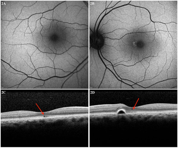
Fig. (2) Case 2: (A) Right eye macula autofluorescence OCT image showing no autofluorescent abnormalities. (B) Left eye macula
autofluorescence OCT image with hyperfluorescence parafoveally. (C) OCT through the right eye fovea showing defects in the inner
segment, outer segment, and inner high reflective layers. Mild hyperreflectivity is noted in the external limiting membrane anterior to the
lesion. (D) OCT through the left eye fovea shows a pigment epithelial detachment parafoveally with no subretinal fluid and disruption of the
inner high reflective layer and the inner and outer segment junction.