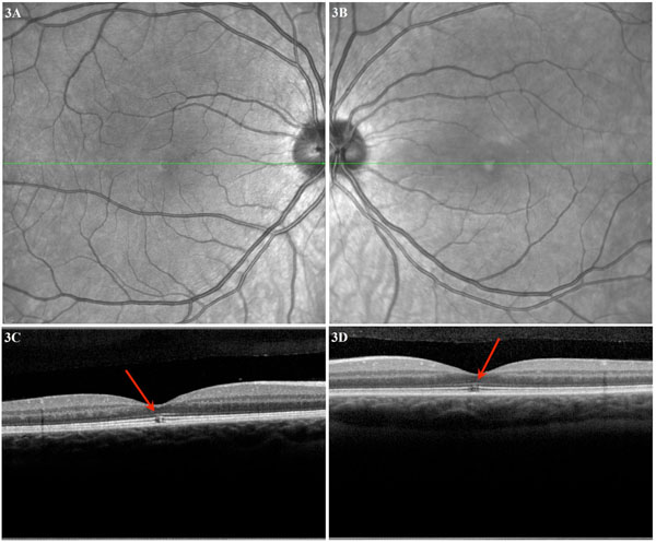
Fig. (3) Case 3: (A) Right eye macula red-free OCT image and (B) left eye macula red-free OCT image showing a brighter foveal area. (C)
OCT through the right eye fovea showing lesions in the inner segment, outer segment, and inner high reflective layers. Note the
hyperreflective external limiting membrane anterior to the lesion. (D) OCT through the left eye fovea showing disruption in the inner
segment, outer segment, and inner high reflective layers.