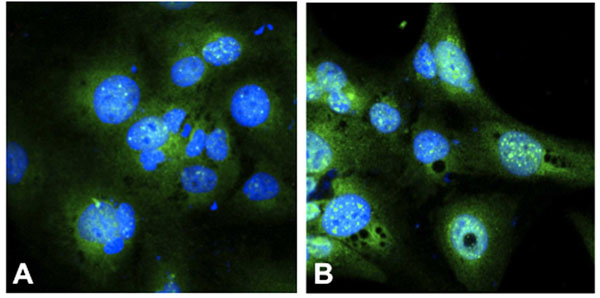
Fig. (4) Immunohistochemistry of the glucocorticoid receptor (GR) showed an increase of GR in the nucleus of (B) dexamethasone exposed
cells (100 µM) as compared to (A) unexposed cells, indicative of translocalisation of the CG-GR complex to the nucleus. The cell nuclei is
stained with Hoechst 33342 (blue), and GR with FITC (green) Original magnification 1000x.