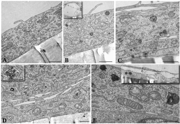
Fig. (5) Morphological changes viewed by transmission electron microscopy. A dose-dependent increase in multivesicular bodies in HLEC
after exposure to 0.1 µM (B), 1 µM (C), 10 µM (D) and 100 µM (E) dexamethasone for 24 hours was evident. Control cells, 0 µM
dexamethasone, are shown in (A). Enlargement of a multivesicular body is shown as an inset picture in (D). Multilayering of cells, present
after 1, 10 and 100 µM dexamethasone exposure, is shown in (C), (D) and (E). Vacuoles also occurred, being more frequent in number and
size with increasing concentration (see inset in picture B and E). Scale bar = 1 µM.