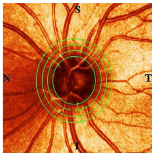
Fig. (1) This picture of a healthy, young individual’s optic nerve
head shows the four concentric ellipses that were superimposed on
each of the participants’ SLT and SLP images. The innermost
ellipse marks the measurements taken exactly on the nerve head’s
rim, the other measurements were taken at 1.25 optic nerve head
diameters (ONHD), at 1.5 ONHD and at 1.75 ONHD. All data
points were saved and documented in the same order: starting on
the temporal side (T) and then proceeding towards the superior (S),
the nasal (N), the inferior (I) segment und coming full circle back to
the temporal periphery.