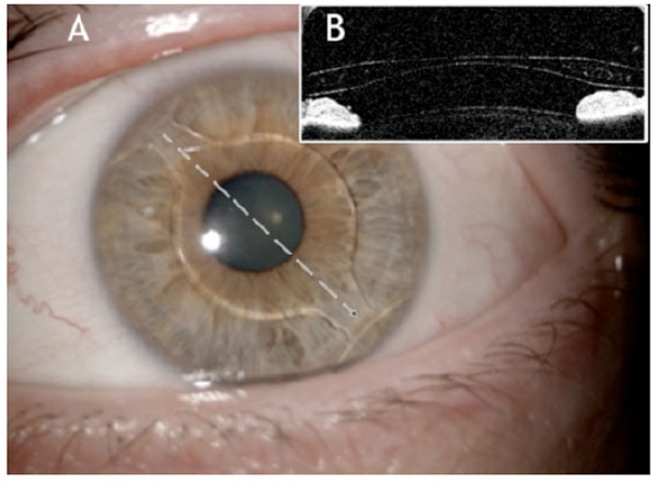
Fig. (1) (A) Slit Lamp image of a Cachet pIOL in the anterior
chamber at 6 months postoperatively. The interrupted arrow shows
the major axis of the lens (60° - 330°). (B) The AS-OCT image of
the pIOL taken along the major axis. Details of the haptic, iris and
anterior surface of the crystalline lens are clearly visible.