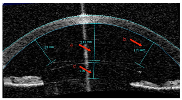
Fig. (2) AS-OCT image of the pIOL at 6-months postoperatively.
The pIOL design allows the maintenance of a virtually safe distance
from the anterior chamber structures, including the anterior surface
of the crystalline lens, the iris, the iridocorneal angle and the
corneal endothelium. The red arrows highlight the minimum
distances, as measured in this study, between (a) the center of the
pIOL and the endothelium; (b) the edge of the pIOL and the
endothelium; (c) the center of the pIOL center and the crystalline
lens.