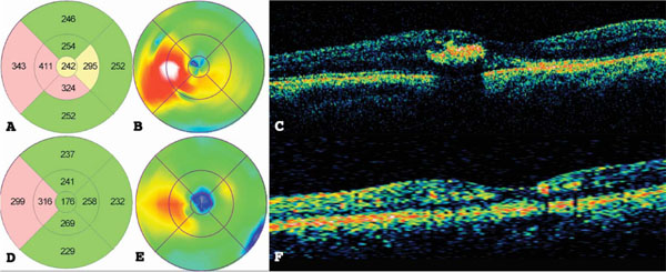
Fig. (2) (A) Macular topographic map, (B) color macular map, and (C) macular cross-section optical coherence tomography (OCT) of the
same study patient from Fig. (1) before treatment. (D) Macular topographic map, (E) color macular map, and (F) macular cross-section OCT
after NAVILAS treatment.

