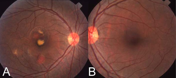
Fig. (1) (A) Color fundus photograph of right eye. Note the large orange subretinal vascular lesions superotemporal and inferior to the center
of the fovea, and serous detachment involving the central fovea. (B) Color fundus photograph of the left eye. Note the lack of any polypoidal
vascular abnormality.