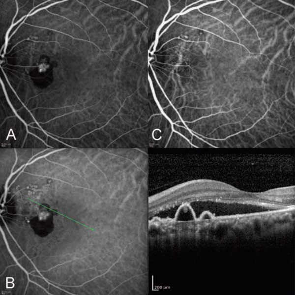
Fig. (3) (A) ICG angiogram of the left eye. Note the new onset of polypoidal subretinal neovascularization. (B) OCT of the left eye
corresponding to the polypoidal complex. Note the subretinal neovascularization lying above Bruch’s membrane and below the retinal
pigment epithelium. (C) ICG angiography after high dose ranibizumab showing resolution of the polyps and RPED with decrease in the
branching vascular network.