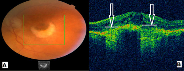
Fig. (1)
A: Color fundus photograph showing an RPE tear. B: OCT of the same patient illustrating clearly the increase of the choroidal depth signal in areas of RPE absence (white arrows).