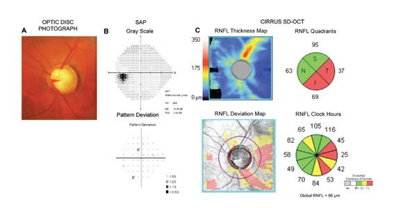
Fig. (4) Example of an eye with preperimetric glaucoma. The optic disc photograph (A) shows diffuse loss of the neuroretinal rim and excavation. However, the standard automated perimetry exam (B) was still within normal limits. Analysis of the retinal nerve fiber layer (RNFL) with SD-OCT (C) reveals extensive loss of the RNFL, compatible with the damage seen on the optic disc photograph.