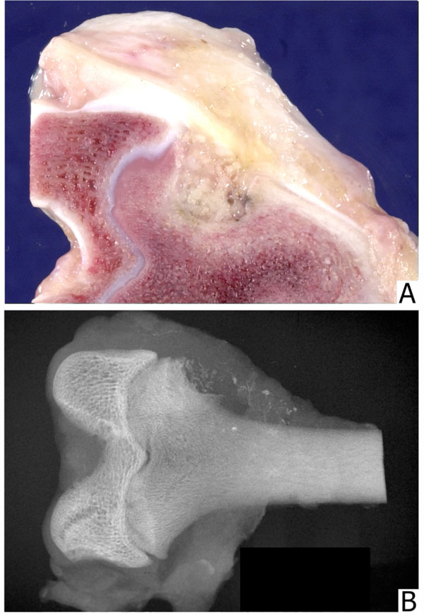
Fig. (4)
(A)
Defect filled with 1-2 mm bone particles; particles are recognizable grossly.
(B)
A radiograph of a defect filled with 800-500 µm particles. The edges of the defect are distinct and the granules are recognizable.