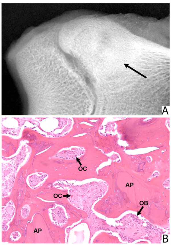
Fig. (5) Defect filled with 300 to 90 µm microparticulate bone allograft. (A) Radiograph shows new bone formation which reaches the epiphyseal line. (arrow) (B) Histologic section from the center of the defect. Allograft particles (AP) are revascularized and are undergoing direct ossification. Both osteoclasts (OC) and osteoblasts (OB) are present. (Stain, hematoxylin and eosin; original magnification, x 100).