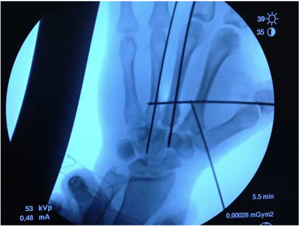
Fig. (3b)
Radiological intra-operative image in which there can be seen the disposition of the Kirschner wires: one of them oblique from the base of the first to the second metacarpal, another transversally and two longitudinally following the axis of the central metacarpals.