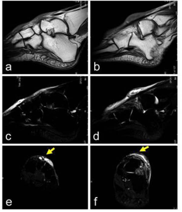
Fig. (1)
A 60-year-old female with a ganglion in the dorsal foot. The cystic lesions, with low signal intensity on a T1-weighted image (a–b) and high signal intensity on T2-weighted images (c–f), are located over the cuneiform to metatarsal bones (a, c, e) and the talus (b, d, f). The dorsalis pedis artery is located under the lesion (yellow arrows; e, f). T2-weighted images with fat-suppression (c-f). Sagittal views (a-d) and axial views (e–f).