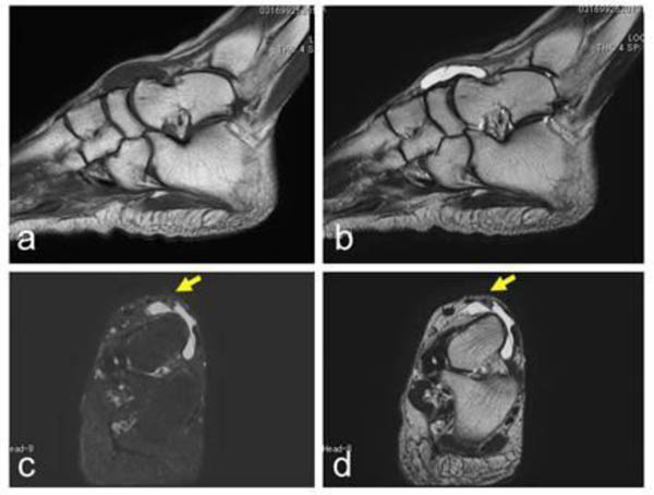
Fig. (2)
A 55-year-old female with a ganglion in the dorsal foot. A cystic lesion, with low signal intensity on a T1-weighted image (a) and high signal intensity on a T2-weighted image (b–d) located over the talus to the navicular (a–b). The dorsalis pedis artery is located over the lesion (yellow arrows; c–d). A T2-weighted image with fat-suppression (c). Sagittal views (a–b) and axial views (c–d).