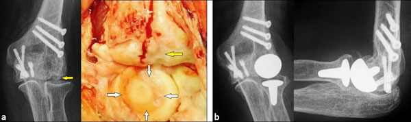
Fig. (4) (Case Presentation): (a) PA radiograph and intraoperative clinical photograph four years after ORIF including one year after partial removal of headless compression screws showing distinctive OA of the capitellum (yellow arrows) without any signs of degenerative changes of radial head radiographically, but intraoperatively there was pronounced deep circular cartilage defect (white arrows). (b) PA and lateral radiographs showing correct positioning and alignment of LRE™ system without any signs of overstuffing.