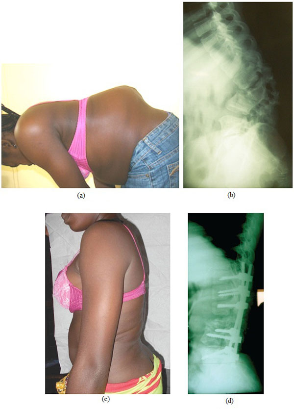
Fig. (1) (a-b). Severe angular lumbar kyphosis involving destruction of L1, L2 and L3. Preoperative x-ray showing a 75° kyphosis.
(c-d). Postoperative clinical and radiographic aspect after correction by pedicle substraction osteotomy and instrumentation from T11 to S1.