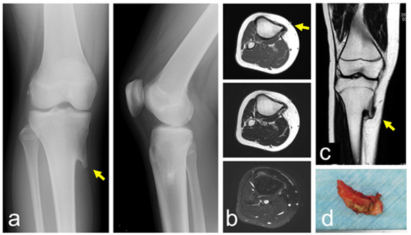
Fig. (2)
A 13-year-old girl with a solitary osteochondroma. Plain radiographs show a lesion protruding from the proximal tibia (a) (left: antero-posterior view, right: lateral view). MRI shows continuity of bone marrow with the lesion in axial (b) (top: T1 weighted image, middle: T2 weighted image, bottom: T2 weighted image with fat-suppression) and coronal section (c). A photograph of a resected specimen (d). (Yellow arrows indicate the tip of the osteochondroma).