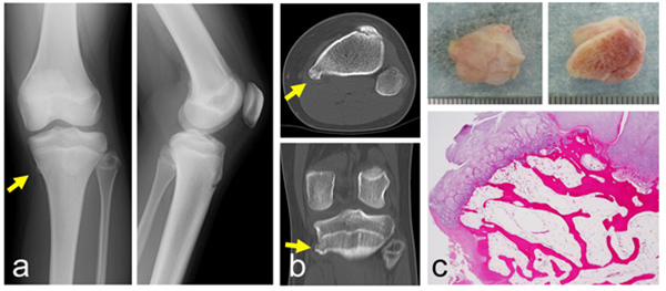
Fig. (3)
A 13-year-old girl with a small solitary osteochondroma. Plain radiographs show a small osseous lesion on the proximal tibia (a) (left: antero-posterior view, right: lateral view). CT shows that the lesion is located at the medial-posterior edge of the proximal tibia (b) (top: axial view, bottom: coronal view). A photograph of a resected specimen from the surface (c: top-left) and the bottom (c: top-right). Histologically, a hyaline cartilaginous cap and underlying lamellar bone trabeculae and fatty marrow are seen (c: bottom). (Yellow arrows indicate the tip of the osteochondroma).