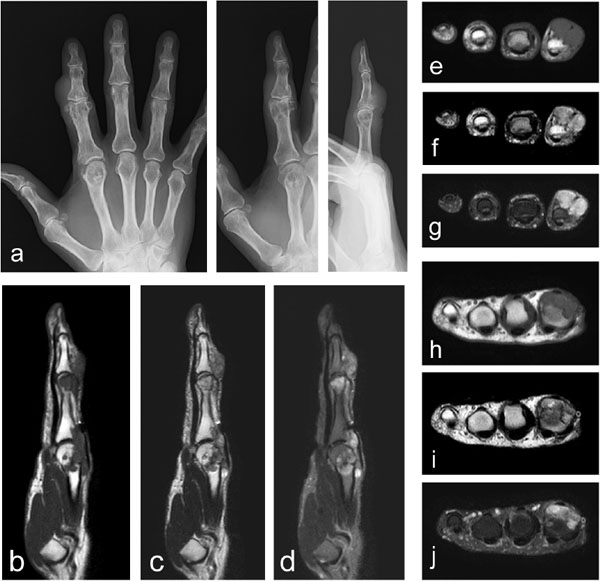
Fig. (1)
Images of a 65-year-old female with extra-articular tenosynovial chondromatosis. The plain radiographs of the index finger of the right hand show irregularity of the cortex at the distal end of the proximal phalanx and of the second metacarpal bone (a: anteroposterior; left, oblique; middle, lateral; right). MRIs (b-j) show multinodular lesions around the proximal interphalangeal joint (e-g) and metacarpophalangeal joint (h–j) levels with homogeneous low signal intensity on the T1-weighted images (b, e, and h), and heterogeneous low to high signal intensity on the T2-weighted images (c, f, and i) with fat-suppression (d, g and j). Osseous invasion is apparent.