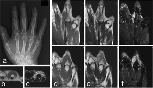
Fig. (4)
Images of a 66-year-old male with extra-articular tenosynovial chondromatosis of the right ring finger. Plain radiographs of the hand showed no abnormality (a). An MRI showing multinodular lesions with homogeneous low signal intensity on the T1-weighted images (b and d) and heterogeneous low to high signal intensity on the T2-weighted images (e) on the dorsal side of the proximal phalanx. Fat suppression on the T2-weighted images demonstrates an extension of the lesion (c and f). The top and bottom images of panel d-f are sequential.