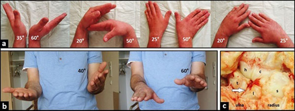
Fig. (2) (Case Presentation, pre- and intraoperative findings): (a) Clinical photographs demonstrating impaired wrist joint motion in comparison to the uninjured right wrist; (b) Clinical photographs demonstrating impaired supination and pronation in comparison to the uninjured right wrist; (c) Intraoperative clinical photograph demonstrating posttraumatic pancarpal wrist joint OA, note the disruption of LTL (arrow) in the absence of disruption of SLL that confirmed the preoperative radiographic finding of PUCT type I.