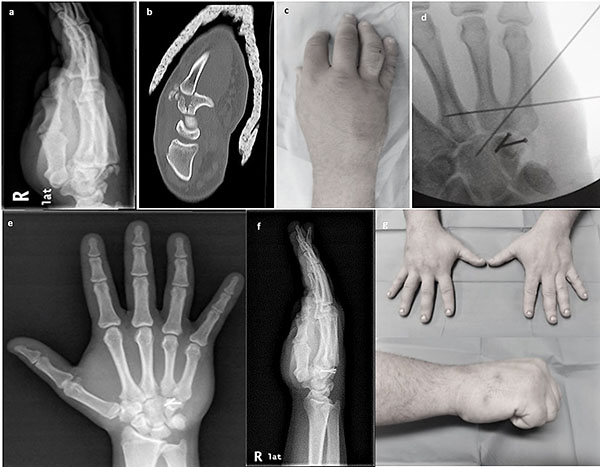
Fig. (1)
(a) Oblique 30o X-ray of the injured wrist showing dislocation of the 4th and 5th metacarpals. (b) CT scan showing the fracture of the hamate body, (c) clinical photo of the wrist showing diffuse swelling over the lesser metacarpals, (d) anteroposterior intraoperative (C-arm) X-ray showing fixation of the hamate with 2 small screws and reduction of the CMC dislocation with 2 KW, (e, f) anteroposterior and oblique X-rays of the wrist at 18 months showing healing of the hamate and congruent hamatometacarpal joint and (g) clinical photos of the wrist showing good range of motion and grip at the latest follow up.