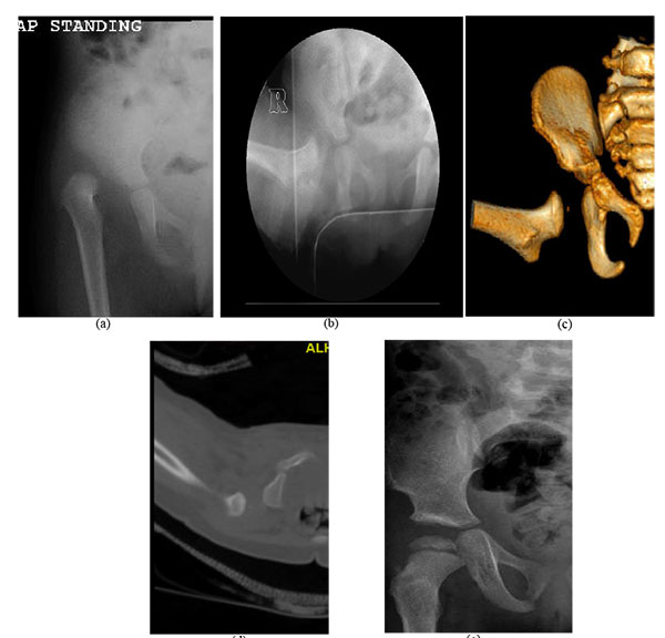
Fig. (2)
Right hip dislocation in an 18-month old girl. (a) Preoperative radiograph. (b) Intraoperative radiograph after closed reduction and cast application. (c) Frontal 3D CT-reconstructed images. (d) Axial cut showing the posterior dislocation. (e) Follow-up radiograph 22 months after salvage with open reduction and Dega osteotomy.