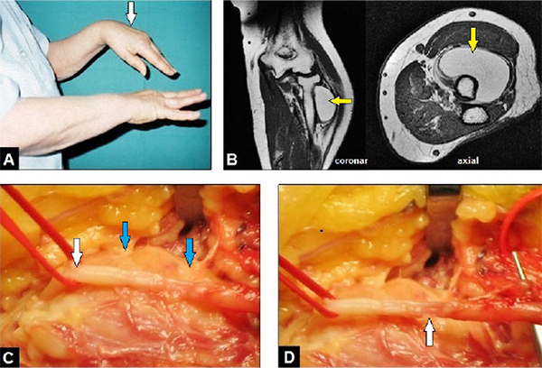
Fig. (1)
(Case presentation, initial and intraoperative findings according to the first procedure): (A) Clinical photograph of both forearms showing left (arrow) complete loss of wrist and thumb's extension and incomplete loss of extension in MCP joints II-V; (B) MRI demonstrating the monstrous GL (arrows) which surrounds the proximal radius shaft approximately a half part of its total circumference; (C) Clinical photograph showing the radial nerve (white arrow) which was fixed to the capsule of the GL (blue arrows); (D) Clinical photograph after monobloc removal of the entire GL and careful dissection of the radial nerve showing the overstretching-related partial disruption of the nerve with a gap of 1 cm involving a half part of its overall circumference (arrow).