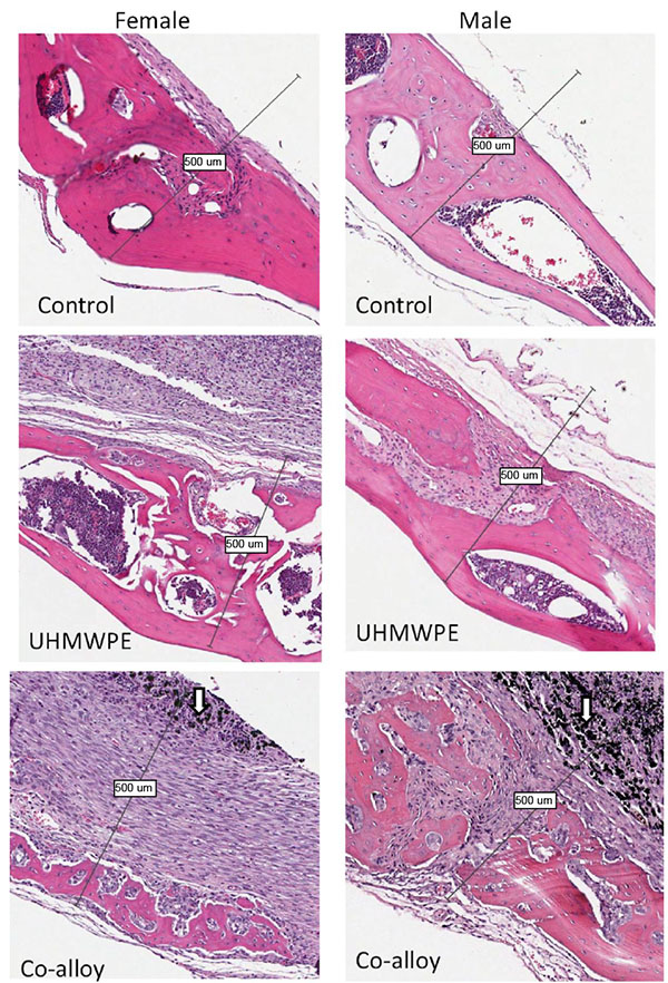
Fig. (4) Histological Analysis of Calvaria Tissue. Examples of histological sections used for histomorphological calculations of bone/osteolysis area of sham, Cobalt-alloy and UHMWPE particle treated calvaria of female and male mice (arrows indicate metal particles) shows the calvaria bone destruction by invasive inflammatory tissue induced by both UHMWPE and CoCrMo particles. Representative images show the relatively larger inflammatory tissue pannus and increased bone resorption associated with CoCrMo when compared to UHMWPE particles (Note: Bars indicate approximately 0.5mm).