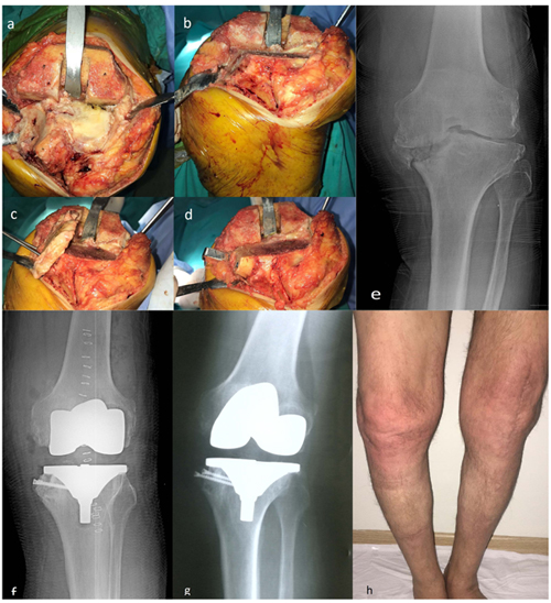
Fig. (1)
Intraoperative photographs of 75 years old patient’s left knee, a; tibia medial plato defect is seen, b; after tibia proximal cut medial side is prepared for fixation, c; tibial cut autograft is fixed, d; autograft is cut, e; preoperative radiographic image, f; postoperative immediate radiographic image, g; first year control visit radiographic image, h; first year photo of lower extremity.