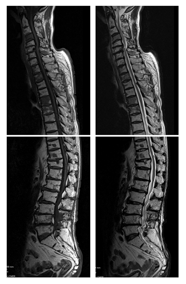
Fig. (2)
Bone marrow reconversion in an 83-year-old male. MRI demonstrates a well-circumscribed diffuse lesion with low signal intensity in the whole spine on the T1 weighted image (left) and high signal intensity on the T2 weighted image (right). The signal intensity is almost the same as that of the spinal cord.