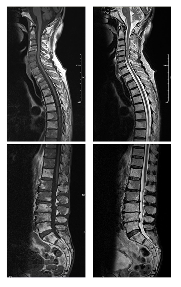
Fig. (4)
Bone marrow reconversion in an 86-year-old male. MRI demonstrates a diffuse lesion with low signal intensity on the T1- weighted image (left) and high signal intensity on the T2-weighted image (right). The signal intensity is almost the same as the spinal cord on the T1-weighed image and slightly higher on the T2 weighted image.