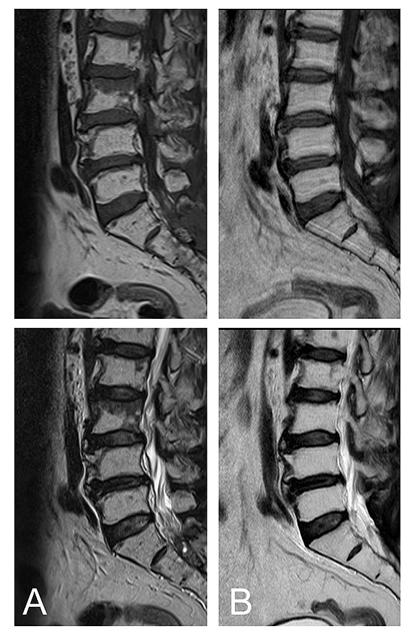
Fig. (6)
Bone marrow reconversion in a 66-year-old male. MRI demonstrates a diffuse lesion with low signal intensity on the T1-weighted image and high signal intensity on the T2-weighted image. After radiation to the posterior portion, the red bone marrow disappeared (12 month interval) (B). (top: T1 weighted image, bottom: T2 weighted image).