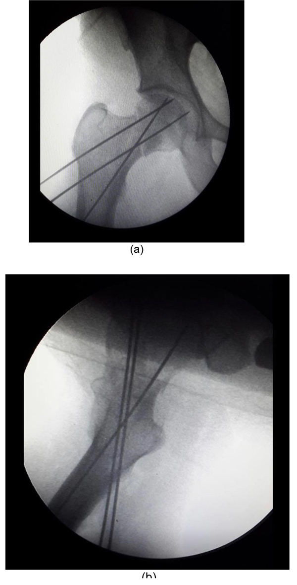
Fig. (4) a) Showing final position of guidewires in the AP view of 76 yrs old female who sustained fracture neck femur. b) Showing final position of guidewires in lateral view with distal guidewire in dorsal oblique plain and proximal and middle guidewires in ventral oblique plain.