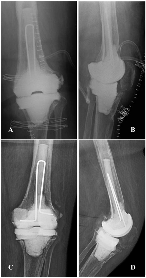
Fig. (2)
X-ray evaluation immediately after the first stage
(A, B)
and a few days before reimplantation
(C, D)
with the spacer in situ. Femoral and tibial bone defects are evident.