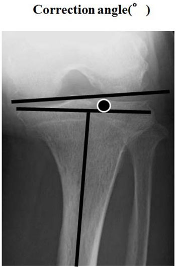
Fig. (4)
Epicondylar view. The correction angle consisted of the posterior condylar surfaces and the perpendicular line of the lower leg axis.