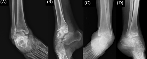
Fig. (3)
Pre-operative X-rays of case I (A, B) and case II (C, D) showing the challenging and severe equinocavovarus deformity.