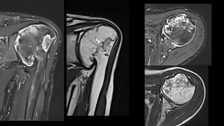
Fig. (3)
Coronal (left) and transverse (right) MRI scans T1 weighted (left and top) and T2 (right and bottom) at age 13.5 years. Hyperintense regions and irregular articular surface of the humeral head and corresponding irregularities of the deformed glenoid.