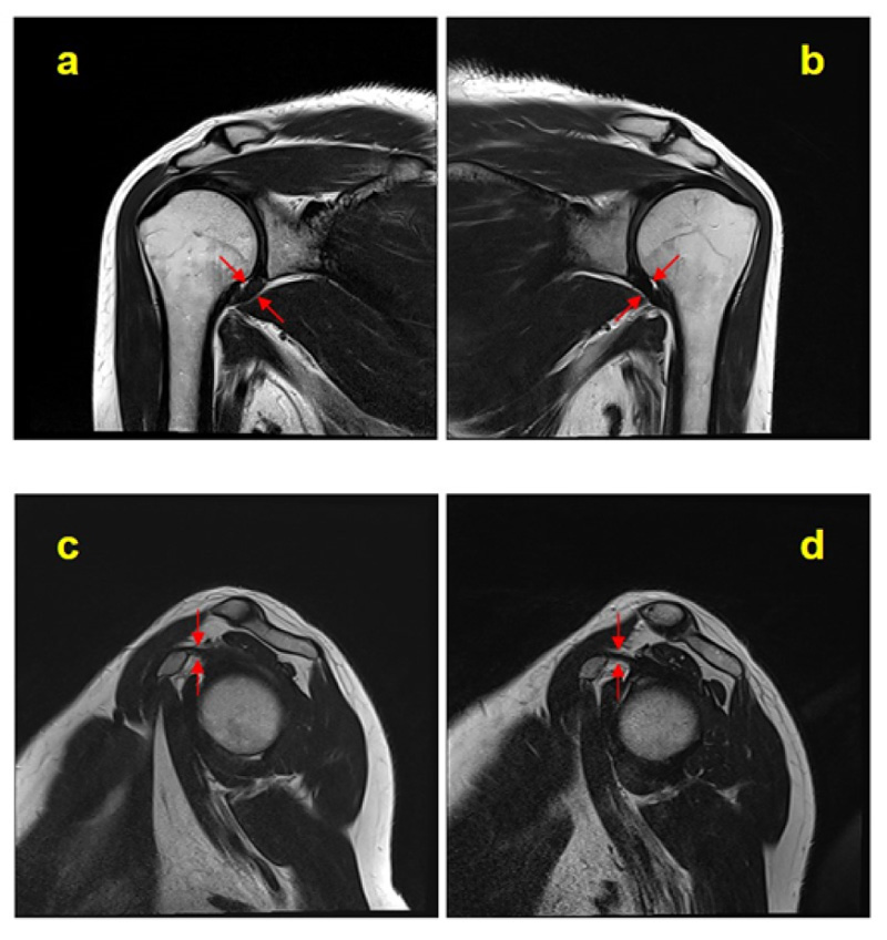
Fig. (1)
Bilateral T2-weighted MR coronal (
a
and
b
) and sagittal (
c
and
d
) images (
a
and
c
is a frozen shoulder and
b
and
d
is a contralateral shoulder). Arrows: measuring sites of the axillary pouch joint capsule and coracohumeral ligament.