Fig. (2)
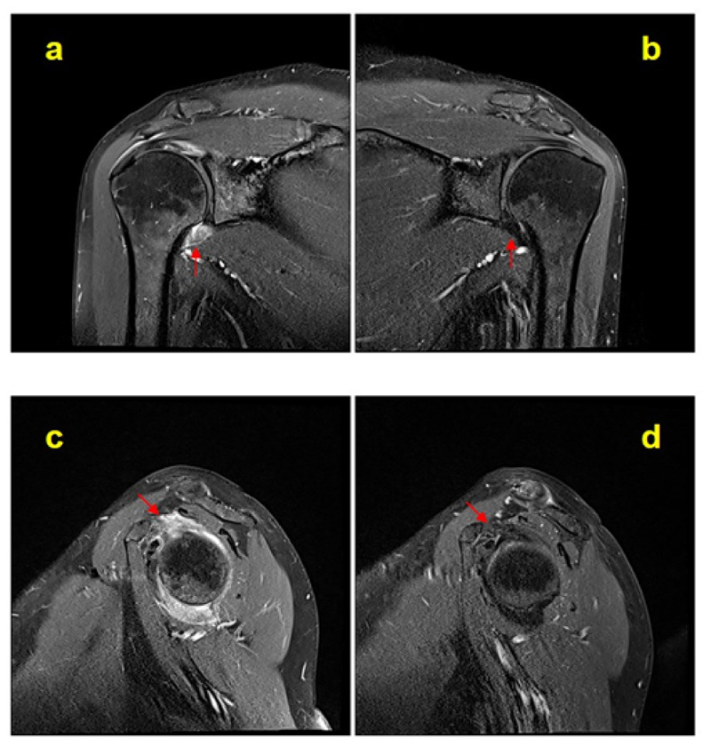
Bilateral contrast-enhanced T1-weighted fat-suppressed MR coronal (
a
and
b
) and sagittal (
c
and
d
) images similar to Fig. (
1
). Arrows indicate measuring sites of the axillary pouch joint capsule and rotator interval enhancement.