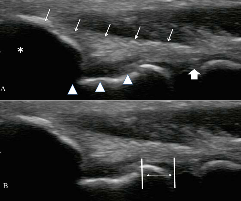
Fig. (2)
Ultrasonographic image of the medial elbow. A. Ultrasonographic images including both the top of the humeral medial epicondyle (asterisk) and the medial tubercular portion of the ulnar coronoid process (large arrow) are displayed simultaneously. The ulnohumeral joint space is observed as an anechoic space between the trochlea (arrowhead) and the coronoid process. The ulnar collateral ligament (UCL) (arrow) is identified as a band-like structure that attached to the medial epicondyle and the tubercular portion of the coronoid process. B. The horizontal distance between the distal-medial corner of the trochlea and the proximal edge of the medial tubercular portion of the coronoid process was measured, and this measurement was considered the medial joint space (MJS) of the elbow (arrows).