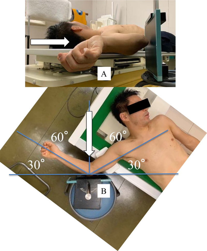
Fig. (3)
Photograph depicting the technique used for valgus stress radiography using gravity. A. Lateral view. Anteroposterior radiographs were obtained as gravity stress was applied to the elbow. The direction of X-ray exposure (arrow) is shown. B. Anteroposterior view. Subjects were placed supine on a table with the shoulder in 90 degrees of abduction, the elbow in 60 degrees of flexion, and the forearm in a neutral position. An elbow flexion angle of 60 degrees was ensured using a triangular polystyrene foam frame set at the correct angle. The direction (arrow) and angle of X-ray exposure are shown.