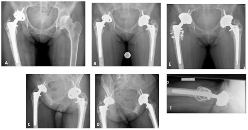
Fig. (3)
Juvenile Idiopathic Arthritis of both hips.A - AP radiograph of the pelvis several years after the right THA. The right hip has been replaced with a cemented stem and a cementless cup with screws (hybrid THA). Note the high placement of the socket due to prior dysplasia. The left hip shows advanced arthritis.B, C, D - Years later, the left hip has been replaced with a hybrid THA. The right hip now shows advanced polyethylene wear and periprosthetic osteolysis.B- AP radiograph of the pelvis; C- Iliac oblique Judet view; D- Obturator oblique Judet viewPost-operative AP (E) and cross table lateral (F) radiographs of the revised right THA. The entire cup was replaced due to its small size and failed locking mechanism. The femoral head size was increased. The stem was well fixed; the deficient calcar was strut grafted.