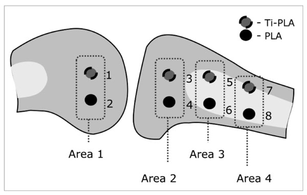
Fig. (3)
Schematic drawing of the implantation sites (1-8) in cancellous bone of the femur (area 1) and cancellous and cortical bone in the proximal tibia (area 2-4). One implant of PLA and Ti-PLA type was inserted in each area.