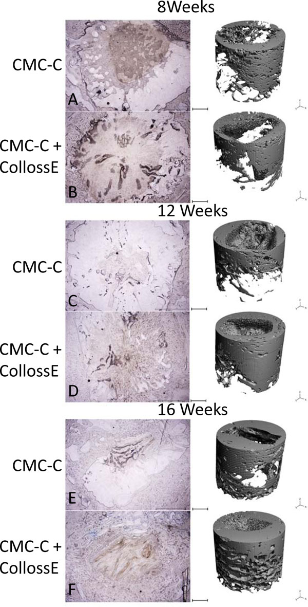
Fig. (5) Comparison of representative histological slices with 3-dimensional reconstructions of the drill defects for control and intervention group at all three time points. In the histological, the bone is colored blue with toluidine blue staining. The images show approximately 80% of the drill hole. The drill hole is easily recognizable with its uncolored marrow space surrounded with blue colored bone. In the CMC-C group, no or little bone formation is present (A), whereas peripheral membranous bone formation is visible in the CMC-C + Colloss E group (B). After 12 weeks, lamellar bone formation was observed at the edges of the defect in the intervention group and woven bone was present throughout the outer parts of the defect (D). No lamellar bone was observed in the control group (C). After 16 weeks, lamellar bone in newly formed trabecular bone structures was visible in the periphery of the intervention group bone defect and new bone was observed almost throughout the defect (F). The trabecular formation was less extensive in the control defect (E). There was no multilocular reossification present at any time point. The 3D reconstruction supports these observations on the right side of the illustration. Bars between the two columns represent 2 mm on the µCT reconstructions and 1,2 mm on the histological images.