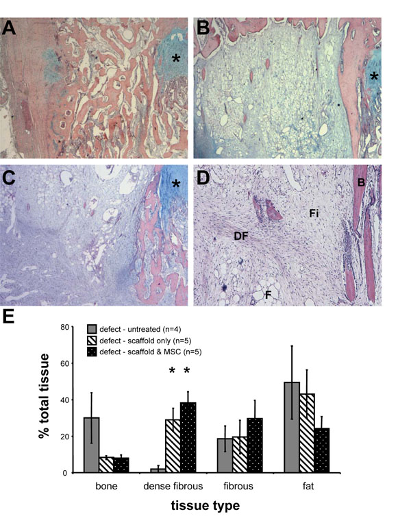
Fig. (3) Surgical creation of a growth plate defect and analysis of repair tissues 5 weeks after untreated or treated with interpositional
transplantation of Gelfoam scaffold alone or seeded with autologous BM MSC. Representative images of repair tissues on cross sections
stained with Haematoxylin, Eosin, and Alcian Blue (A-C). The location of the growth plate is indicated by *. A representative example of
extensive bone bridge formation is observed in the untreated defect (2x) (A). A representative example of growth plate defect treated with
Gelfoam scaffold only (2x) (B), and defect treated with Gelfoam scaffold and autologous MSC (2x) (C). An example of different tissues
observed within defect (4x) (D), which include bone (B), fat (F), fibrous tissue (Fi), and dense fibrous tissue (DF). Using Image Analysis
software, the proportions of each repair tissue type were determined and are presented at the % mean area ± SEM of the total injury area for
each group (E). Statistical significance between treatment groups is indicated by a * (Mann-Whitney, p < 0.05).