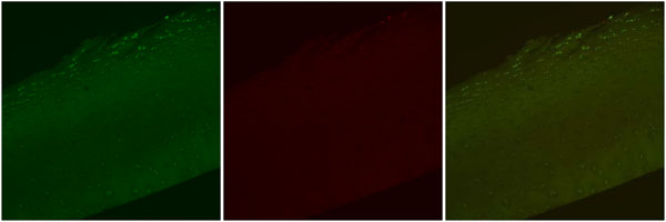
Fig. (2) Confocal laser microscopy images, Control. Representative images depicting Live cell stain (green), Dead cell stain (red), and a
combined image with both Live cell and Dead cell stain. Note the fibrillation within the Superficial Zone and the normal appearing elongated
chondrocytes both within and around the fibrillation. Dead cells are observed only in extruded positions. Original magnification 10x.