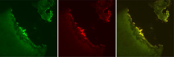
Fig. (3) Confocal laser microscopy images, Glider. Representative images depicting Live cell stain (green), Dead cell stain (red), and a
combined image with both Live cell and Dead cell stain. Note the level of tissue loss which brings the post-treatment residual necrotic
cartilage layer into the Transitional Zone. The Transitional Zone typically demonstrates spherical chondrocytes of less dense population than
the Superficial Zone. Original magnification 10x.