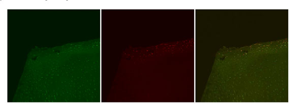
Fig. (4) Confocal laser microscopy images, Paragon. Representative images depicting Live cell stain (green), Dead cell stain (red), and a
combined image with both Live cell and Dead cell stain. Note that the Superficial Zone remains present, but with significant dead cells
observed uniformly throughout this zone and into part of the Transitional Zone. Original magnification 10x.