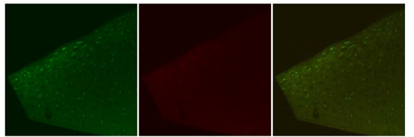
Fig. (5) Confocal laser microscopy images, Ceruleau. Representative images depicting Live cell stain (green), Dead cell stain (red), and a
combined image with both Live cell and Dead cell stain. Note the decreased distance of live cells to the surface. These cells exhibit elongated
morphology typical of the Superficial Zone. Dead cells are not observed in either the Superficial or Transitional Zone. Original magnification
10x.