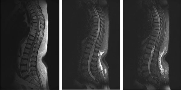
Fig. (1) Magnetic resonance tomography examination, presenting from left to right: T1-sequence median sagittal, T2- sequence median
sagittal and paramedian sagittal layers. High signal intensity is visible at the level of the 12th thoracic and the 1st lumbar vertebra, surrounded
by diffuse edema extending cranial and caudal.